There are some very important structures linked to the construction of a memory. The thalamus is actively engaged in directing attention. The caudate nucleus is often described to be part in the formation of memories of instinctive skills. The mamillary body has been referred to episodic memories in scientific papers. However, the putamen has a decisive function when it comes to procedural skills. Emotions linked to memories can be found and associated with parts of the amygdala. However, one of the most named structures when it comes to the formation of memories is the hippocampus in the inferior temporal lobe. Other parts of the temporal lobe are actively engaged in holding general knowledge.According to Carter, 2009, “the cerebellum is associated with conditioned memories“ (2009, p.154). The parietal lobe as a whole is often described to have an associative function when it comes to visuospatial memories. Lastly, the frontal lobe is the main region of interest when it comes to processes of the working memory. It is not yet answered if there are other structures which are engaged in the formation of memories. Nevertheless, an insight into the types of memories is necessary for the understanding of the injury of patient S.S.
The formation of a memory starts with an experience that is intensive enough in order to be encoded as a memory or, to be more precise as an engram, which is the simultaneous firing of neural networks that make up the memory. According to Carter, 2019, it takes two years to consolidate the changes that create along-term memory but, once encoded, that memory may remain available for life. Furthermore, “long-term memories include events from a persons life (episodic memories) and impersonal facts (semantic memories)“ (Carter, 2019, p.158).
Most literature as well as research in the field of memory and learning is based on a model as shown in figure 1. The human memory system is divided into a declarative and a non-declarative branch. The declarative branch is further divided into semantic and episodic memory. However, the non-declarative branch is divided into 4 subtypes , namely, the procedural skill, the conditioning, a non- associative and a priming subtype.

Figure 1: Organization of the memory (Purves et al.,2013)
Another model presents the formation process of memory. It must be mentioned that all memory formation is based on sensory input which is stored shortly, around one to two seconds, in a sensory register until it is further processed in the human short-term memory. Here, a storage of information is dependent on the different items. Seven plus minus two items can be stored for around 15-30 seconds. Via rehearsal and retrieval, a consolidation in the long-term memory is most likely. The central executive, or working memory is a combination of a phonological loop, an episodic buffer and a visuospatial working memory. It results in higher order brain functions and managing processes as well as organizing, planning, executing and decision-making processes.

Figure 2: memory consolidation model (Bear et al., 2016)
The aforementioned types of memories depend the crucial cellular and molecular mechanism of long-term potentiation. According to Hebb´s Law, “cells that fire together wire together“. Applied to the formation of memory, cell assemblies that fire simultaneously are seen to represent memories at an internal cortical level which are activated by an external stimulus. Due to repeated firing from similar neurons, these connections strengthen and an engram for a specific external stimuli develops. Even after a period of time and a partial activation of such an engram the whole internal representation is represented (Bear et al., 2016).
At the level of a synapse, many different processes take place. One of them is an increase in neurotransmitter release into the synaptic cleft due to a higher firing rate. Another change is the development of more receptors at the level of the postsynaptic dendrite. These two changes result in a stronger depolarization which is known as long-term potentiation. There are two types of long term potentiation, the early long-term potentiation, measurable until one to two hours, and late long-term potentiation. Chemically, this can be explained through the development of AMPA receptors which are present due to cascades of enzymes building phosphatases which lead to further AMPA expression and an increase in the permeability of neurotransmitter. All in all, a higher sensitivity for glutamate arises.
Attention is one of the most essential neurobiological function that allows humans to constantly and dynamically choose between important and also interesting stimuli in the internal and external environment on which greater neural processing resources can be directed for more intense examination. “When researchers talk about attention in the literature, the term „modulation“ is frequently used“ (Andrewes, 2001, p.140). Modulation itself is often used to explain a more efficient way of doing a task and can similarly be used to explain an increase in a specific performance. A deficit in attention modulation is most commonly diagnosed with ADHD or ADD, although there are other pathophysiological and neuropsychological disorders which are of less interest.
Hierarchically, attention is one of the key cognitive processes of the human brain and constantly in interaction with many other cognitive processes. Several attention models have been described in the past to explain the process of attention. All of these models suggest that there is a limited attention capacity, especially when “interference is increased above a certain level and the task requirements are increased“ (Andrewes, 2001, p. 141). This Phenomenon can be observed when two different tasks need attentive resources in order to perform both of them. As a result, an inefficiency in both tasks will be visible. Some Neuroscientists, Neuropsychologists, and researchers as well as medical professionals from any branch might argue that perceiving the internal and external environment might be more important, it can be stated that attention and perception are dependent from each other. On the one hand, a stimuli that receives no attention will remain invisible in any environment. On he other hand, remaining in an attentive state without a stimuli, no focus can be established on anything. Indeed, in activities of the everyday living perception and attention go hand in hand and are vital for adequate performance. Attention is often misinterpreted and mixed with general states of cortical arousal. The main relationship between attention and arousal is that an awake medium arousal stage is supports effective attentiveness. Other stages of arousal are low and high arousal where attention in more ineffective. Another “key distinction between arousal and attention is that attention can be selectively focused“ (Purves et al., 2008, p.250). Explaining attention dates back into the 19th century, where William James, a neuroscientist famous for the James-Lange theory in the field of emotion stated, that “attention is the taking possession of the mind in clear and vivid form, of one out of what seem several simultaneously possible objects or trains of thought. It implies withdrawal from some things in order to deal effectively with others“ (Purves et al., 2008, p. 251). Attention, as one of the cognitive processes of the brain, can be directed towards any external and internal stimuli. External stimuli belong to the visual and auditory as well as olfactory sensory system.

Figure 3: Types of attention (Andrewes, 2001, p.147)
Internal stimuli are those from skin receptors in the skin, proprioceptors at the neuromuscular junction and those belonging to the organization of the human homeostasis. However, it lies in human nature that attention is directed to auditory and visual stimuli predominantly and rapidly. As already mentioned, attention is mostly present with other cognitive processes. Therefore, it must be mentioned that three independent but cooperating attention networks can be observed. The reticular system is predominantly active to set the body into an alert state. It is also known as the first system of arousal. It serves as a surveillance network. From here, two other attention networks must be divided. One the one hand, the posterior attention system (PAS) and on the other hand the anterior attention system (AAS). In order to master any task-oriented behavior, the PAS and the AAS work closely together. As these systems are highly intertwined, a neuroanatomical view on the attention networks is needed to point out which cortical structures are responsible for attention processes.
When it comes to attention and functional as well as structural explanations, the cocktail party effect is often described to explain the phenomenon of attention. “This term refers to the situations in which multiple conversations and other sounds are occurring and simultaneously but a listener can selectively focus on one voice or conversation and effectively turn out the others“ (Purves, 2015, p. 251). This effect belongs to those of the auditory attention system but can similarly applied to visual attention.
In general, a dorsal and a ventral fronto-parietal attention system can be highlighted when it comes to attention process from a resting state brain, which is the default mode network. Recent studies highlight the fact that neither of the aforementioned attention systems work isolated and that “the flexible interaction between both systems enables the dynamic control of attention in relation to top-down goals and bottom-up sensory stimulation“ (Vossel et al., 2013, p.1). The Ventral fronto-parietal attention network is linked to bottom-up attention and from a functional point of view, bottom-up attention is called exogenous attention whereas the top-down attention is also called endogenous attention. The illustration below shows the ventral fronto-parietal attention network and the two different pathways for top-down and bottom-up attention are highlighted. In general, this frontal-parietal attention network “is thought to modulate activity in the sensory cortices and other brain regions, resulting in more effective processing of some inputs and less processing of others“ (Purves et al., 2019, p.640).

Figure 4: Top down attention (Bear et al.,2016)

Figure 5: Bottom-up attention (Bear et al.,2016)
A huge amount of research has been done so far concerning the exploration of the attention networks. It is no coincidence that most of the research that has been done shows a decisive integration of the visual system e.g. many researchers have shown until today that the dorsal and the ventral stream are crucial for the perception of space and therefore influence attention processes (Kolb et al., .2015, p. 613).
Romeas and others (2015), presented significant results in 3D MOT training and decision-making processes in soccer players by looking at essential skills in soccer players. In their study they focused on dynamic visual acuity. Christian Vater (2019) and Vater and others (2017) pointed out how selective attention affects the detection of motion changes within the peripheral vision system. Moreover, the multiple object tracking method was applied as intervention in his study. Besides the visual system, starting with a light stimulus eye at the retina until it is finally processed in the visual cortex, other brain regions have a close relation to the the visual cortex (Harris et al. 2020). An important brain
region that is closely related to attention processes is the frontal eye field.

Figure 6. Activation of attention networks due to a visual stimuli (Haines et al., 2017)
This area produces saccadic eye movements in the contralateral eye. Inevitably, its location is near to the premotor areas of the brain. Moreover, the frontal eye field has a general connection to “numerous areas known to be influenced by attention, including areas V2, V3, V4, MT, and the parietal cortex“ (Bear et al. 2016, p.735). These areas closely interact with a brain structure in the midbrain called the superior colliculi. These structures, together with parts of the thalamus, more precisely, the pulvinar
nucleus and the lateral geniculate nucleus, form main structures of cortical attention networks.

Figure 7. The dorsal and the ventral stream (Vickers, 2007)
Most commonly known are the dorsal and ventral visual streams, of which the ventral stream is connected to the question what is seen whereas the dorsal stream is connected to locations of objects (Vickers, 2007). The following figure shows the neuroanatomical location of both streams.
The dorsal stream runs along the parietal lobe whereas the ventral lobe runs along the temporal lobe (Sheth & Young, 2016). Interestingly, the positions P3 and P4 on the 10-20 system directly measures activity in these dorsal regions. This region is called the lateral intraparietal cortex and plays a decisive role in directing eye movements coupled with attention as well as processing of visuospatial orientation. Therefore, activity in the LIP should be visible during an attention task in an EEG of a test subject. Furthermore, appropriate processing of visual information results in good reaction times.
Different types of lesions that affect the medial temporal lobe as well as some diencephalic and prosencephalic structures might lead to amnesia. There are two main types of amnesias that can be differentiated. Anterograde amnesia affects the memory formation after the injury whereas retrograde amnesia affects the memory prior to the injury. Furthermore, a transient global amnesia can be diagnosed which affects the memory more temporally and belongs to amnestic syndromes, of which three other types (Korsakoff-syndrome, mild cognitive impairment and hysterical amnesia) can be classified. An injury to the prefrontal cortex might result in deficits in the working memory. The working memory is inevitable in order to master any mental task based on the central executive functions as mentioned in figure 2. in section 1.2. However, forgetting memory belongs to the essential mechanisms in humans as not every aspect of past experience must be stored to guide some of the personal future actions.
Moreover, it is necessary to create general plans from specific actions after learning e.g. to open a bottle of water where it is important to know that the cap must be rotated into left direction similarly to other bottles to get open.
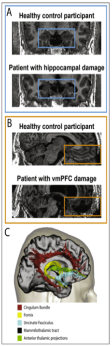
Figure 8: Hippocampal and pfc damage (McCormick et al., 2018)
Five frequency bands (Delta 0-3Hz, Theta, 4-7Hz, Alpha, 8-13Hz, Beta,14-30Hz and Gamma, >30Hz) can be observed via electroencephalography (EEG) within the brain which result from cortical activity from pyramidal cells measured at the scalp of a subject. To be more precise, a summed potential is measured, amplified and visualized in a specific software. The following chart shows the different frequency bands and the morphology that can be measured.
In an active awake state, alpha activity in the posterior part of the brain (occipital) is the most common one in healthy people. Furthermore, theta and beta waves are found in an awake state. This situation changes under stressful situations, in an alert mode as well as in drowsy state. To sum up, “the different frequency bands reflect the electric operations of the brain“ (Demos, 2019, p. 58). In situations, when focus and concentration is needed, alpha and beta waves are most prominent. Therefore, these frequency bands will be further explained in the following.
Alpha waves are the first waves that have been described in neuroscience in 1924 as a result of the work of Hans Berger. Therefore, Alpha waves are often also called Berger waves. In a calm and peaceful state, alpha waves are present in the normal healthy adult brain. There is a predominance in the posterior, occipital part of the brain. Alpha waves are divided into two separate ranges, slow alpha waves (8-10 Hz) and fast alpha waves (10-12 Hz). One of the most important findings according to alpha waves is the alpha blocking when the eyes are closed. Immediately, alpha amplitude increases and show a special morphology called alpha spindles. When reopening the eyes, a reactivation of the idling visual system causes a decrease in alpha waves. An increase in alpha waves with eyes open while doing any task can be linked to the alpha inversion problem. This phenomenon is of relevance when it comes to EEG measurements in this research topic. Alpha waves are not treated separately from another wave form called MU waves when it comes to QEEG brain mapping which have the same eight to twelve Hz range. They can be differentiated from normal awake alpha rhythm by looking at the raw EEG data concentrating at the morphological differences at the measurements from the electrodes C3 and C4.
Beta is frequently seen in alert or attentive mode. Beta is also “defined as fast wave“ and “it has been associated with being focussed, analytical, externally oriented or in a state of relaxed thinking“ (Demos, 2019, p.79). Weak beta power may also be an EEG marker (signature) for poor cognitive performance. High beta (20-35 Hz) is linked to peak performance and other cognitive processes. Most commonly associated with peak performance and cognitive processing are high Beta waves (20-35 Hz). EEG measurement is mainly used “to support clinical diagnosis or symptoms“ (Demos, 2019, p.88). As a result, not much is reported and provided about special normal distribution, e.g. the frequency bands during attention processes. The mere activation of a brain area, e.g. the prefrontal cortex does not finally prove the assumption that only attention processes are present.
Figure 9. The international 10-20 system (Demos, 2019)
Recent research in the field of neurophysiology as well as literature concerning Neurofeedback has shown that complex networks are responsible for processing memory such as the theta/beta ratio which will be analyzed in patient S.S. QEEG as well. According to study material in the education of Neurofeedback therapists, a good theta/beta ratio is 1:2. That means a 7 hertz theta frequency paired with a 14 hertz beta frequency is a marker for better learning and memory skills. In a QEEG this will be presented in the section power ratios.
The following pathophysiological diseases were found in patient S. S. in July 2015. A rupture of the anterior communicating artery, an organic psycho syndrome, an amnesic syndrome and hydrocephalus malresoptivus. In addition, the installation of a VP shunt was ordered. Furthermore, a bifrontal hemicraniectomy with right cranioplasm was conducted. During the hospital stay, the patient suffered a pulmonary embolism and symptomatic epilepsy.
The treatment concept in the first months after the injury was carried out as an inpatient in a neuro-cognitive special ward. This included the treatments cognitive disorders after brain damage of various causes such as head trauma, strokes, hypoxia, inflammatory processes, metabolic disorders, tumors and neurodegenerative diseases. In the case of patient SS, work was explicitly done on memory performance and orientation. The impairments of attention and language as well as the self-structuring of everyday life of the ability to act and behavior were also trained. The aim of the rehabilitation stay was to improve cognitive abilities, to promote everyday skills, to stabilize existing skills, to install everyday structures, to achieve independence in everyday life and to gain personal responsibility. The goal was communicated to promote quality of life as well as participation in social life. An interdisciplinary treatment team consisted of nurses, occupational therapists, physiotherapists, language therapists, social workers, neuropsychologists, sports therapists and doctors.
Patient S. S. is always accompanied by her mother and her father to the follow-up examinations. Physically she has recovered well from the subarachnoid hemorrhage. At the time of the experiment, she can cycle longer distances again and is quite independent in everyday life. Patient SS is allowed to drive again when accompanied. However, there are still severe disorders of attention, concentration and memory so that reintegration into the labor market has not yet been possible. In the clinical examination, the scars are free of irritation; however, there are many places where the skin is drawn in the area of bone gaps. Bone necrosis was excluded and there was no need for neurosurgical action at the time of the experiment.
The following figure points out the different changes in personality based on the aforementioned disorders. McCormick et al. describe in their research paper „Comparing and Contrasting the Cognitive Effects of Hippocampal and Ventromedial Prefrontal Cortex Damage: A Review of Human Lesion Studies“ the effects of lesions similar to those of patient S.S. on personality. In the following chapter, the scientific background as well as a justification for this study will be explained.
Before the start of the planned experiment, a research paper search was carried out. The database Pubmed was used to seek for research in the field of amnesia, Neurofeedback, virtual reality and attention in the context of rehabilitation that was already carried out. The following search strings were entered into the database to find adequate research.
- Visual attention and learning and amnesia – 146 results
- Visual attention and memory and amnesia – 166 results
- Visual attention and memory and amnesia and VR – 0 results
- Neurofeedback and amnesia – 7 results
- Neurofeedback and short-term memory – 40 results
- Neurofeedback and short-term memory and attention – 21 results
- Virtual reality and amnesia – 12 results
- Virtual reality and attention – 923 results
- Virtual reality and attention and memory – 172 results
- Virtual reality and attention and memory and rehabilitation – 77 results
- Virtual reality and attention and memory and rehabilitation and training – 48 results
The following studies were found eligible for a deeper examination and helpful information was extracted. McCormick (2018) and others, compared cognitive effects based on the region of cortical lesion in either the ventromedial prefrontal cortex or the hippocampus. Their findings lead to precise results concerning personality traits and the underlying lesion. Richard Allen (2018) published an article on recent advances in understanding amnesia with the conclusion that “debate continues regarding the patterns of preservation and impairment across a range of abilities, including semantic memory and learning, delayed recognition, working memory, and imagination.“ (Allen,2018, p.1)
Lin and others (2016), highlighted the use of computerized visual speed of processing training in patients with amnestic mild cognitive impairment. They found significant improvements in attention and memory processing. Larson and others (2011), confirmed in their study that the use of virtual reality is “well-tolerated and engaging and that they could be beneficial for inpatients with severe TBI.“ (Larson et al. 2011, p.1) Zhang et al, 2013 concluded that VR is beneficial as well as valid and reliable.
Based on the theoretical background, the following hypotheses can be deduced and will be tested.
- H0 = No effect of EEG-biofeedback on the QEEG measurement in patient S.S.
H1 = EEG-biofeedback affects the QEEG in Patient S.S.
2) H0 = Virtual Reality has no effect on patient S.S. visual field attention.
H1 = Virtual Reality interventions with patient S.S. results in a better reaction time in the visual field attention test
3) H0 = Upper and lower standard deviations in z-score training does not decrease in 15 trials of EEG-Biofeedback
H1 = Standard deviations in EEG-biofeedback z-score training decreases after 15 trials
4)H0= Virtual reality training with beat saber has no effect on the working memory
H1= Virtual reality increases the working memory based on the digit span test
Based on these hypothesis, the dependent and independent variables can be listed as seen in the figure below.
Table 1
Dependent and independent variables
Subject | Independent variable I | Dependent variable I | Dependent variable II | Dependent variable III |
Patient S.S. | 15 sessions of EEG-Biofeedback z-score training |
Digit span test |
Visual field attention |
Romberg stance |
Virtual reality Training | Digit span test | Visual field attention | Romberg stance |

Figure 10: Phases of the study project
EEG measurements
To measure brain activity in the experimental group, the brain master discovery is being used. It is a 19-channel amplifier and uses the software brain avatar for visualization of brain waves. Usually, this system is used for EEG Neurofeedback. Therefore, it is linked to the online service QEEG pro for converting raw EEG files into a summed QEEG. The EEG cap is a medium free cap. Exemplarily, the following figure shows the QEEG outcome of one of the test subject from which results will be drawn.

Figure 11: Example of QEEG Z-score analyses
Neurofeedback intervention
The intervention used on the patient is based on Z scores on all 19 channels. The zokul values indicate the percentage of the z-scores of patient S.S. that are within the z-values. This changes dynamically when the different frequencies develop towards the normal value. The standard deviation from the normal value of the Z scores can be set and adjusted over the course of the interventions. As part of this intervention, the positive and negative standard deviations were reduced to +2. and -2. Auditory feedback was chosen as feedback when the frequency benders are within these areas. The music played inside the range and the music stopped outside the ranch. Each intervention with the EG Bio Feedback lasted 20 minutes with a preparation time of 15 minutes for the adjustment of the electrodes on the head skin. The feedback is shown as a percentage of reward. There are different approaches to this in the context of neuro feedback therapy. In this intervention, the private was used as a criterion for better neural connections and was therefore not actively influenced. This influence can be determined by increasing or decreasing the limit values of the standard deviation. A dimmer function of the screen could have been used as a further reward. However, this was not done in this intervention
The visual field attention test
The company brain metrics offers several software in the field of neuropsychology, of which one software is the visual field attention test which tests visual attention by measuring the reaction time to a given stimulus in one of the four visual quadrants. Most effectively, it can be used in patients with hemineglect and other types of brain injuries resulting in visual attention deficits. When starting this software some parameters could be set prior to the testing. One of the settings can be either a random set of stimuli or a record of stimuli that are manually entered for a more precise measurement if a hemineglect is known in a patient. Furthermore, it is important to set the distance from which the stimuli are presented on a rather large screen or just at a smaller screen like a notebook screen. It is proposed to choose a distance between 1 and 14 inches. It is also possible to vary the stimuli in this software by setting the pause time between two stimuli preventing from possible learning effects due to rhythmicity. last but not least, a stimulus presentation time can be set for either long or very short presentation of the stimuli which also can be set. By clicking the start button, reaction time is measured as the test subject has to click whenever a stimuli that is presented is perceived. For this test, 50 stimuli were presented to the subject. Figure 9 shows the result screen that is presented for analyzation. The reaction times in the central and peripheral vision as well as left and right eye reaction time will be extracted and compared.

Figure 12. Result section of the visual field attention test
Memory Tap Brain application
The Tab Brain program is based on the neuropsychological test of the DJ Spam Test. A 3×4 large field similar to a chess board in the colors red, blue, yellow and green appears when you start the program. The program randomly presents a field that lights up and at the same time gives the time of appearance in the form of a number in chronological order. This must then be pressed by the test person. It should be noted that in addition to the glowing light, there is a piano sound. One after the other, the program selects another field which the test person has to type in the same order. As soon as the test person can no longer repeat the specified color combination, the number of fields typed is recorded.

Figure 13: Tap Brain test
Romberg Test
The romberg test, named after the german neurologist Moritz Heinrich Romberg, is a neurological examination of the general stance of a patient. The patient has to stand with both feet alined in front of one another. The hands are raised in front of the body and the eyes must be closed. The examiner supervises the sway of the body. The patient is not allowed to orient oneself towards sources of light or sound. A positive romberg test is recorded when the patient can not hold the stance with closed eyes. The patient is supported to not fall over to one side. A strong sway to one side might represent a damage of the vestibular organ on the ipsilateral side. This is also visible while the eyes of the patient are open.
Virtual Reality system and software for the intervention
The HTC Vive pro Eye system was used to carry out the experiment. For this purpose, the system was set up in an appropriate room with sufficient space. The two sensors for creating the virtual room were placed opposite each other at a distance of 4 m. In the settings of steam VR, the software from HTC Vive, only standing position was selected and set instead of the room-filling display. A gaming PC from the brand Aorus with an Nvidia graphics card model HTC 1660i was used to display the virtual world. All content was presented so fluently that the phenomenon of motion sickness could not occur.
Beat Saber Software
The Beat saber software can be described as follows: blue blocks on the right and red blocks on the left approach the person standing in the virtual space. Directional information in the form of arrows can be seen on the blocks in white. The blocks must be pierced with the swords that the test person holds in his hand in the direction of the directional information. It should be said that the swords are also shown in the colors blue for right and red for left. Another essential aspect is that breaking the blocks appears in time with the music. This creates the impression that the smashing of the red and blue blocks is rhythmically linked. A sequence of correctly smashed blocks increases the streak. An error leads to a restart of the counting mechanism. Each song can be played on the levels easy, normal, difficult, expert. For this experiment, the three levels of simple normal and difficult were selected. The highest streaks as well as the total number of broken blocks were included in the data collection.

Figure 14: In-game view of Beat Saber
The living room of patient S.S. was chosen for the experiment. This room provided enough space to carry out the intervention in virtual reality. Furthermore, the EEG could be set up at a table. All existing materials remained untouched in this room and the experimental setup was not changed.


